Faculty/Staff Photography, 1st Place. Ritwick Ghosh. Forest conservation often puts wildlife protection over the interests of tribal communities who live in and around dense forests. For tribal groups to effectively assert their rights, they must establish and strengthen village-level institutions. This image aims to capture the mundane but critical documentation and bureaucracy that are key to upholding tribal groups' legitimacy and power — democratizing environmental governance. The research highlights the land conflicts involved in forest conservation, as well as the many ways communities are fighting to realize their dreams of alternate futures for humans and nature.
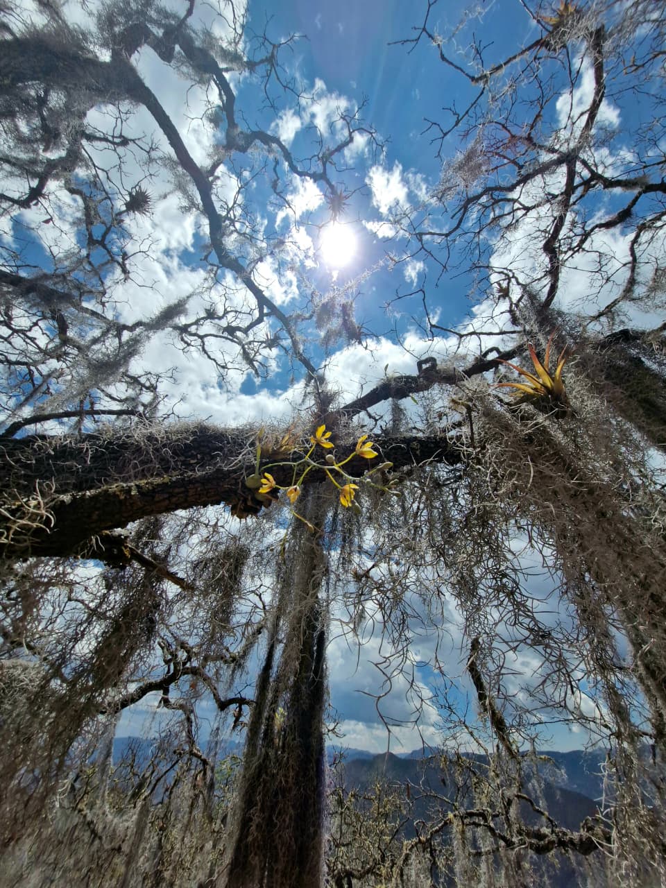
Faculty/Staff Graphics, 1st Place. Marsha Gordon and Robin Vuchnich. As the naturalist and philosopher Henry David Thoreau observed, we all need “the tonic of wildness” for our collective survival as well as to nourish our spirits. This image is a botanical portrait composed of pressed and preserved plant petals, leaves and stems — gathered from the Gregg Museum of Art’s pollinator garden as an adjunct to the interdisciplinary “In Search of Thoreau’s Flowers” exhibition at the Gregg Museum — which combined art and science to reflect on environmental change at a turning point in Earth’s history.Graduate Student/Postdoc Photography, 1st Place. Laura Arango. In the temperate forests of Oaxaca, Mexico, oak trees create canopy spaces that support a rich diversity of epiphytes. This image shows how sunlight filters through the branches, and bromeliads and orchids bring this hidden world to life, weaving a radiant tapestry of color and biodiversity across the forest canopy. The research — which is being conducted in the community of Santa Marías Jaltianguis, Sierra Norte, where common land-use practices shape the forest structure — explores epiphyte diversity across three habitat types: patch cuts, buffer zones and conservation areas, to understand how their presence and patterns change across the landscape.
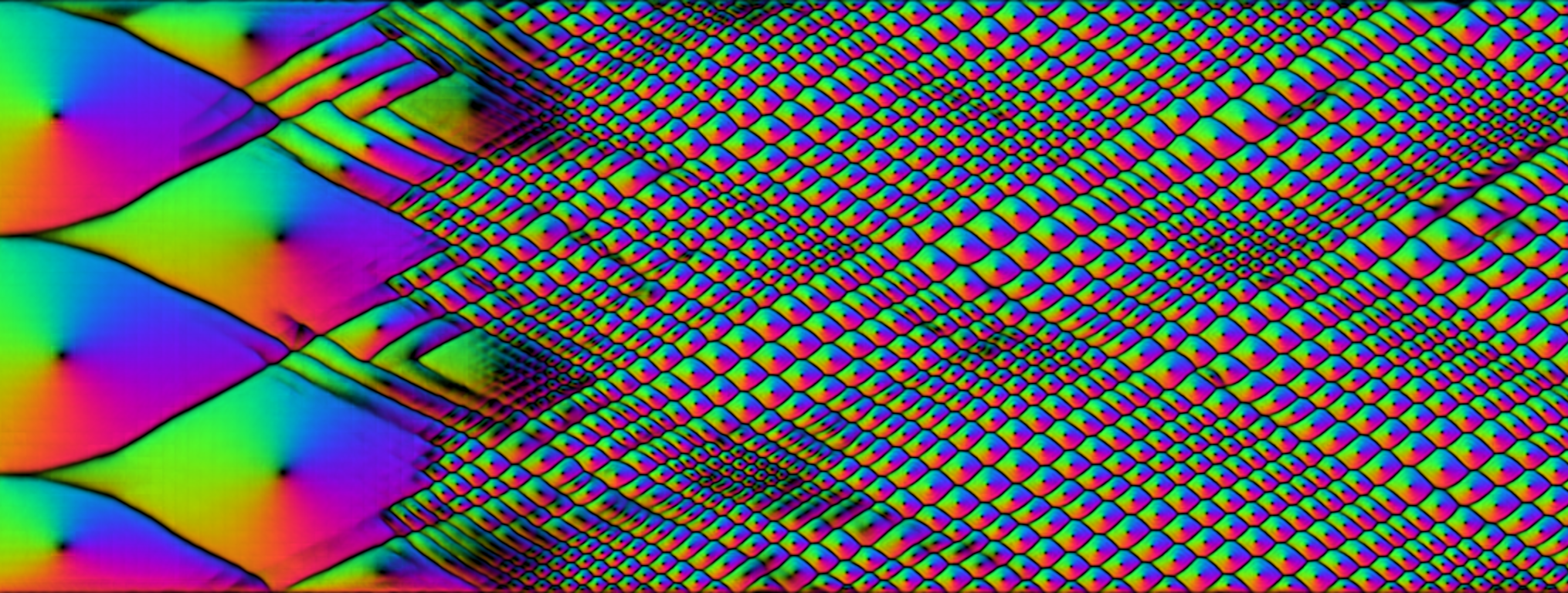
Graduate Student/Postdoc Graphics, Honorable Mention. Jacob Leff. Cellpose-SAM is a version of the popular Cellpose image segmentation framework — a deep learning model trained specifically to identify and outline cells in microscopy images — that integrates Segment Anything Model (SAM) to improve segmentation of cells and other biological structures. The image above depicts the “flows” that the Cellpose-SAM convolutional neural network uses to automatically differentiate and tag cellular structures in the growth and formation of hydrocarbon detonation. This result is post-processed from a 2D computational fluid dynamics simulation in an 8-centimeter channel, with the wave propagating from left to right. Cellpose-SAM allows gaseous detonations to be studied in a faster, more reproducible way, which is useful for modeling propulsion and safety systems.
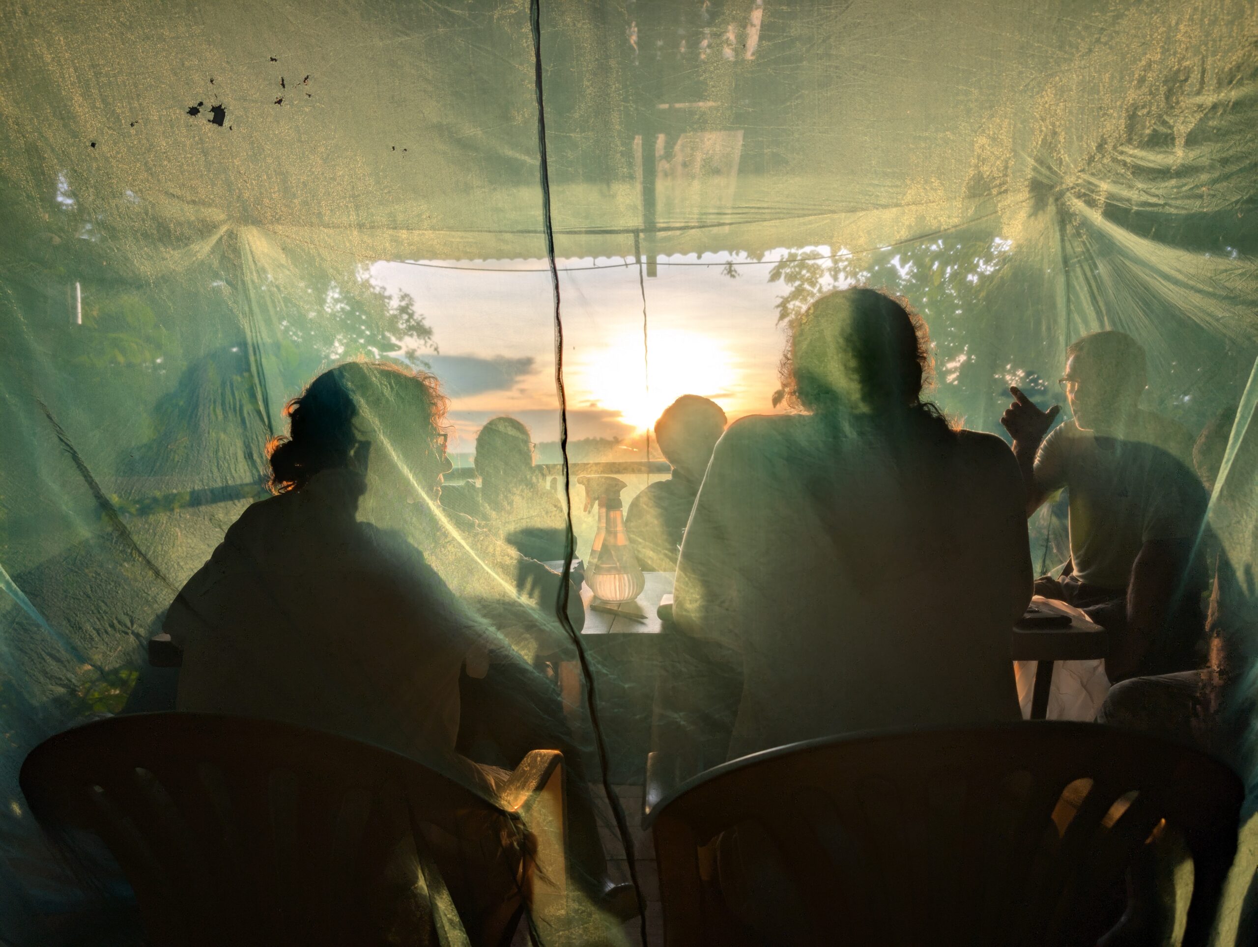
Graduate Student/Postdoc Photography, 2nd Place. Kenneth Geisert. The Amazon rainforest faces increased threats from deforestation, climate change and habitat degradation. Researchers in professor Gareth Powell’s Insect Evolution Lab, in collaboration with the Alliance for a Sustainable Amazon, conducted research spanning multiple projects at Finca Las Piedras Research Station in Peru — aimed at understanding the natural world through the lens of insects. Over the course of a week, researchers set up non-invasive insect traps (shown above) to collect molecular-grade specimens for genetic and morphological analyses.Graduate Student/Postdoc Microscopy, 1st Place. Stephen Colleran. This photo might look like a piece of abstract art, but it’s actually an image of some of the thinnest materials in the world. These two-dimensional materials are created with merely a substrate, bulk crystal and some magic tape that can be found in any classroom. The flakes in this image are about 60 nanometers thick — or 1,000 times thinner than a human hair.
Graduate Student/Postdoc Graphics, Honorable Mention. Alexandria Brown. Through image analysis techniques, root systems can be isolated from the background to quantify growth patterns, branching structure and overall morphology. This image captures the root architecture of a sweet potato slip, digitally processed to highlight its structural form. The root silhouette happens to resemble someone dancing — an unexpected artistic moment emerging from scientific data. While visually striking, the image represents ongoing research focused on understanding how propagation conditions influence root uniformity and development. By analyzing root structure across treatments, this work supports improved propagation practices and more consistent plant establishment.
Faculty/Staff Photography, Honorable Mention. Madaris Serrano Perez. Soil microbiomes, communities of diverse microorganisms living in the soil, are drivers of soil health — and studying them could open the door to more sustainable agricultural practices. The image above shows a researcher collecting samples at a soybean field in Beaufort, NC, in order to study agricultural soil microbiomes. In Perez’s extension program and research lab, a variety of projects are underway to look into microbial diversity as it relates to agricultural practices and the changing environment.Graduate Student/Postdoc Photography, Honorable Mention. Julia Brzezicki. Habitat loss and fragmentation, combined with high elephant densities, increase competition for resources with nearby human populations — which leads to conflict. This research focuses on understanding wild elephant behavior and genetics, particularly the factors that influence human-elephant conflict. The photograph shows Brzezicki collecting behavioral data on adult Asian elephants in Minneriya National Park in Sri Lanka. Conducted in collaboration with the Sri Lanka Elephant Project, Brzezicki’s research aims to inform local mitigation strategies by identifying why some elephants are more likely to crop raid than others and to promote coexistence between elephants and humans.
Faculty/Staff Photography, 2nd Place. Heather Frantz. Uncovering the details of parasite life cycles, particularly with specimens collected over several decades, allows scientists to better understand current and historical ecosystem health. This image shows Ph.D. candidate Alex Nelson searching for freshwater snails on floating vegetation in Yates Millpond. Nelson doesn’t study the snails themselves; rather, the research centers on trematode (flatworm) parasites that use snails as first intermediate hosts — before going on to infect vertebrate species such as snakes. In the lab, Nelson investigates which parasite species infect which snake species by dissecting roadkill and preserved snakes from the North Carolina Museum of Natural Sciences.
Graduate Student/Postdoc Microscopy, 2nd Place. Byeunggon Kim. Society needs sustainable solutions for removing microplastics from our waterways in order to protect the health of global aquatic ecosystems — and the safety of drinking water. This electron microscope image shows a round microplastic particle trapped by dendritic particles with architected structures that have tree-like branches, which act as microscopic webs. Using natural forces, the branches wrap around and firmly capture the plastic. By applying advanced materials science to environmental cleanup, these advanced microscopic nets offer a powerful way to filter out pollutants and protect our ecosystems — helping preserve biodiversity and cleaner oceans for future generations.Graduate Student/Postdoc Microscopy, Honorable Mention. Carlos Morales. Aspergillus niger, a common, widespread fungus, causes certain fruits and vegetables to grow black mold. This image shows Aspergillus niger under a microscope, growing from infected sweet potato tissue. Recovering the same fungus from this lesion helped confirm it caused the disease — turning an invisible plant infection into visible proof. This research supported efforts to protect roots in storage and reduce losses for Ugandan farmers by helping identify the fungus responsible for sweet potato rot.
Undergraduate Student Graphics, 1st Place. Fiona Wolfe-Roberts. Physics can help us understand and appreciate nature’s seemingly mysterious patterns. In this image, two stars bound by gravity orbit each other in the binary system Vela-X1 — and a neutron star two times the mass of the Sun rips matter off its supergiant companion. The research graphic models the supergiant’s atmosphere as it leaks mass, causing powerful velocity flows.
Faculty/Staff Microscopy, Honorable Mention. Liv Borges. Streblospio benedicti is a common coastal species of plankton that can now be found throughout the world. The species is often used as a bioindicator to assess the health of its environment. This image shows planktotrophic Streblospio benedicti larvae stained with an antibody to assess where chitin — shown in red — has developed. Undergraduate Student Microscopy, Honorable Mention. Audrey Johnson. Previous research into the crucial neurodevelopmental protein cyfip2 has shown that mutants without functioning cyfip2 are hyperactive to auditory stimuli compared to fish with functioning cyfip2 (wild-type fish). This image depicts a zebrafish model used for a study focused on improving our understanding of the auditory system. The aim of this research is to investigate where in the auditory pathway differences occur, building upon basic science principles of the auditory system.
Graduate Student/Postdoc Graphics, Honorable Mention. Alp Tezbasaran. Inside a boiling reactor or a fuel injector, the boundary where liquid ends and vapor begins is constantly shifting — which makes tracking directly every bubble and droplet impossible. This image visualizes a workaround called the “level-set” method, which builds a continuous mathematical field across the domain — a smooth landscape that quietly carries every surface inside it. From this single map, computers simulate boiling, atomization and the breath of an ocean wave. Each glowing ring is the boundary of an individual bubble or droplet; the horizontal seam splits vapor (top) from liquid (bottom) across the whole scene. The simulation recovers all interfaces in one clean stroke, without ever drawing them. Radiating bands trace distance to the nearest surface: closer means brighter, farther sinks toward dark.
Graduate Student/Postdoc Photography, Honorable Mention. Zehta Fazler. CRISPR-Cas9 allows geneticists to make precise, targeted changes to a plant’s DNA. This research focuses on growing and selecting maize plants edited using CRISPR. In the field, researchers manually pollinate plants in order to prevent the spread of edited genes to other plants. This picture shows a graduate student hand-pollinating gene-edited maize. By studying how specific genes influence plant performance, the research aims to support breeding efforts that produce reliable yields while minimizing environmental impact — to advance a more sustainable future in agriculture.Graduate Student/Postdoc Microscopy, Honorable Mention. Mohammed Kayes Patoary. Nature is the ultimate architect, crafting everything from skeletal structures to intricate shells. This micrograph captures crystal patterns grown using a method that mimics these very biological processes. The research explores how calcium-based minerals can be guided to grow in controlled ways, much like the formation of bones (calcium phosphate) and shells (calcium carbonate). By maintaining minerals in a unique, liquid-like state, it’s possible to direct their growth before they harden into their final form.
Graduate Student/Postdoc Photography, Honorable Mention. Sierra McMurry. To improve conservation efforts, researchers study the relationships animals have with their main sources of food. This image, taken with a camera trap, shows a red-tailed hawk catching a garter snake. Using both camera and seed traps, McMurry investigates how tree seeds like acorns and pinecones impact mammal communities.
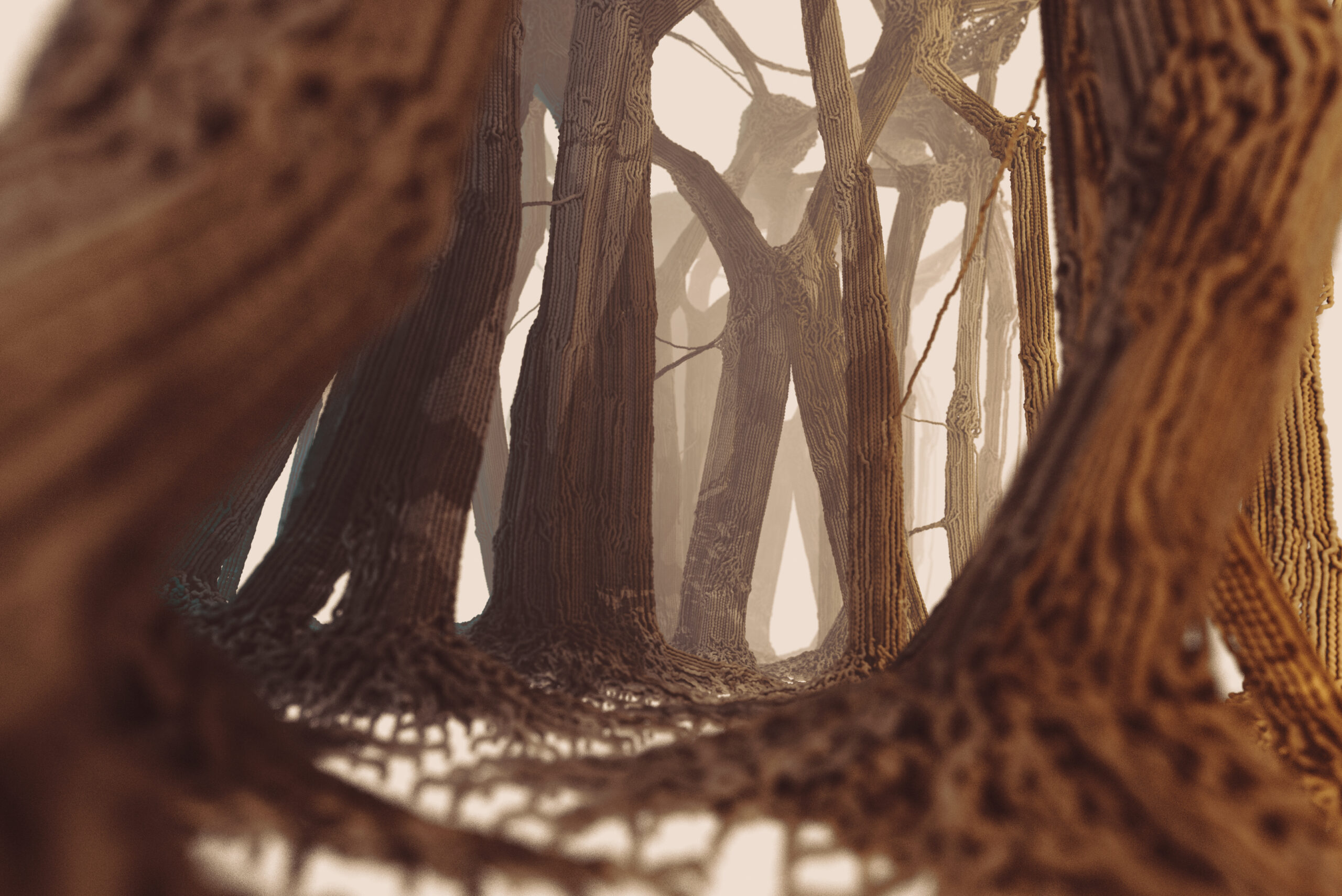
Undergraduate Student Photography, 1st Place. Brock Davis. Students taking a general entomology course (ENT 425) at NC State were given an assignment to photograph and document 25 families and eight orders of insects. This image shows one of the favorite insects Davis photographed and documented — the Differential Grasshopper.Faculty/Staff Photography, Honorable Mention. Abolhassan Pishahang. Living systems can contribute to adaptive, responsive design. Pictured above is a bio-fabricated scaffold — developed at the NC State Crafts Center — designed to support the growth of living material within a computationally generated structure. The scaffold acts both as a structural framework and as a medium for cultivation, enabling the emergence of bio-composite surfaces over time. This research demonstrates how design can move beyond static materials toward dynamic systems that integrate biological growth as an active component. Developed for bio-lighting applications, the porous lattice was fabricated to host organic matter, allowing seeds to germinate and develop directly within the form.
Faculty/Staff Microscopy, 2nd Place. Yasaman Ghaemi. To figure out how neurological diseases begin, it’s important to first understand how brain cells grow and organize during early development. This image captures neurons and glial cells, including astrocytes, weaving together to form the three-dimensional structure of the mouse brain cortex. The elegant shapes and interactions create a dynamic cellular landscape — and what emerges is a vivid mosaic of life, where each cell contributes to the beauty and complexity of the brain. In the image above, an immunostained brain section is magnified under a microscope using light-sheet fluorescence. By revealing processes at the single-cell level, the research could support new treatments and improve brain health.
Graduate Student/Postdoc Graphics, 2nd Place. Elias Zauscher. Water distribution systems deliver treated water to residential, commercial and industrial consumers. To better serve their population base, though, water utilities need system models —which can be costly and time-consuming to create. This image shows a small part of the training dataset the researchers used to develop AI methods for generating distribution network models. Graduate Student/Postdoc Photography, Honorable Mention. Sarah Cochran-Murray. A recent anthracnose disease outbreak has North Carolina pepper growers concerned. Since its first sighting in 2024, reports of the disease have tripled. Colletotrichum scovillei — a new, aggressive fungus — is spreading throughout pepper fields across the state. The new species of anthracnose disease (pictured above) can cause heavy yield losses because it infects young crops and thrives in spite of modern management strategies.
Undergraduate Student Photography, 2nd Place. Gabrielle Schoeffel. When investigating how different knitted jacquard fabrics compare in appearance and size when made using different machine settings, the researchers found that even when the same size was programmed, fabric samples often came out in different shapes and dimensions. They ran many tests to learn how fabric structure affects final size and appearance. However, rather than discard the abundance of extra samples used during testing, the researchers transformed them into a quilt (pictured above) — which visually represents the research in a creative way.
Undergraduate Student Microscopy, 1st Place. Sarah Siegle. Corals may inhabit less than 1% of the ocean floor — yet they’re an essential source of food and habitat for roughly 25% of all marine species. Unfortunately, 30-50% of coral reefs have already been lost — and many more are at risk due to climate change, pollution, habitat destruction and ocean acidification. This image of the species Orbicella faveolata shows bright clusters of potential coral-associated microbial aggregates, which potentially house Endozoicomonas. The globally distributed bacteria Endozoicomonas — which has the potential to strengthen coral immune systems, cycle important nutrients, and help corals respond to stress — is the focus of Siegle’s research.Undergraduate Student Microscopy, 2nd Place. Elijah Cornatzer. Granulomatous Meningoencephalomyelitis (GME) is an incurable, often fatal inflammatory disease that occurs in the brains of dogs. This image shows the Hippocampal section from a canine diagnosed with GME. The cause of GME is not fully understood, so this research looked for characteristics seen in more widely understood brain diseases by using established markers that identify hallmark pathology for these diseases. The image above shows a sample in which the researchers looked for markers associated with Alzheimer’s disease by staining for Tau (a protein associated with AD) and Astrocytes (a cell associated with neuronal inflammation).
Graduate Student/Postdoc Photography, Honorable Mention. Arindam Mandal. In the field, research isn’t always about big machines and spreadsheets; often, it’s about the quiet, microscopic battles happening right under our nose. This picture of a caterpillar exemplifies a constant, if uninvited, companion in daily scouting. While the researchers’ focus is on soil physics and plant health, these tiny residents serve as a reminder that agriculture is a living, breathing ecosystem.
Graduate Student/Postdoc Microscopy, Honorable Mention. Fatima Ezzahra Allam. Cellulose, derived from natural sources like plants, can organize itself at the nanoscale to form intricate and highly ordered patterns. This image reveals the microscopic structure of a thin film made from cellulose nanocrystals combined with a polymer called Jeffamine. By mixing in Jeffamine, the researchers created a flexible material in which these tiny building blocks interact and rearrange — producing unique textures observed under microscopy. Nanoscale arrangements play a crucial role in determining a material’s properties, such as strength, flexibility, and responsiveness to the environment. This work explores how sustainable, bio-based substances can be engineered at very small scales to create advanced functional materials.Undergraduate Student Microscopy, Honorable Mention. Thomas Sutton. Cells precisely control gene expression by regulating messenger RNA, the molecule that carries genetic information from DNA to produce proteins. This research specifically focuses on a group of RNA-binding proteins that coordinate these complex regulatory networks — and how these mechanisms can be harnessed to develop new therapeutics. Using the model organism Caenorhabditis elegans, the researchers examined the molecular pathways involved in translation initiation, a fundamental process that also occurs in human cells. The image above shows target proteins tagged with fluorescent markers using CRISPR-Cas9 genome editing to visualize the RNA-binding proteins’ activity in living organisms — offering a fascinating window into the cell’s inner workings.
Graduate Student/Postdoc Graphics, 1st Place. Sergei Rigin. This image depicts the simulation of a magnetic nanoparticle coated with flexible polymer chains. When exposed to a rotating magnetic field, the nanoparticle spins — causing the attached polymer layer to swirl, twist and wrap around itself like a tiny tornado at the nanoscale.
Undergraduate Student Microscopy, Honorable Mention. Malcolm Albergo-Radisch. The human body is at peak regenerative capacity during embryonic development, which is governed by a complex interplay of molecular signals. This image, taken at 30 days of gestation, shows progenitor cells (visualized in green) precisely localized within the annulus fibrosus — a ring that’s responsible for the mechanical integrity of an intervertebral disc. Utilizing a gene-edited pig model, an ideal translational bridge to human biology, the researchers mapped the spatial distribution of LGR5+ cells within a developing intervertebral disc. This work provides the biological framework necessary to develop targeted therapies for intervertebral disc repair.Faculty/Staff Microscopy, Honorable Mention. Rebeca Castro. The kidney’s job is to filter waste from our blood, but in tubulointerstitial nephropathy, that system begins to break down. This image shows a paper-thin slice of a diseased kidney, stained with a special dye to reveal its inner workings in vivid color. The colors tell the story: red highlights healthy, functioning tissue, while cyan marks areas overtaken by scar tissue, a sign the kidney is losing the battle. Scattered throughout are tiny, round structures called glomeruli, the kidney’s microscopic filters, where clusters of red blood cells are visibly trapped mid-filtration. The stark contrast between the thriving red regions and the spreading cyan creates a vivid map of a kidney slowly losing function — and makes the damage impossible to ignore.
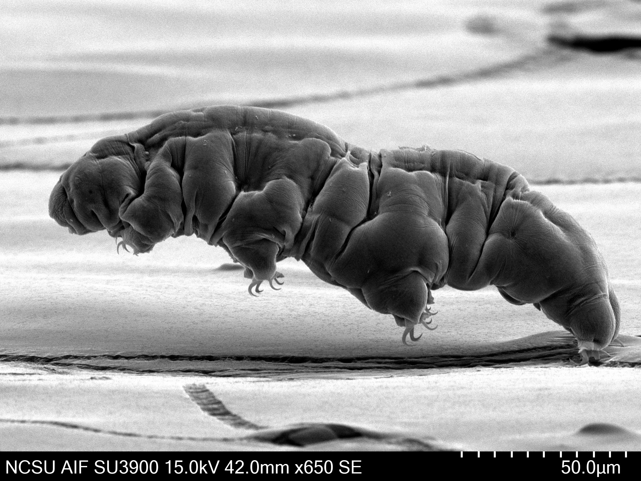
Faculty/Staff Microscopy, 1st Place. Aaron Bell. About 1,500 species of tardigrades — which are among the most resilient organisms known to science — can thrive in most of the diverse ecologies found in Earth’s biosphere. This image shows a tardigrade galloping, magnified under a microscope. Tardigrades can survive severe conditions, such as exposure to extreme temperatures, air deprivation, radiation, dehydration, extremely high and low pressures, and starvation. Tardigrades have even survived exposure to outer space.
Graduate Student/Postdoc Video, 1st Place. Faby Claure. This video shows a lab test using a shake table to simulate the effects of an earthquake on a bridge column. By recreating real earthquake motions, the researchers study how bridges behave, where damage occurs, and how to better predict this damage. In this test, the column is pushed to the point of collapse — allowing the researchers to observe failure mechanisms and improve the design of safer, more resilient bridges.
Faculty/Staff Video, 1st Place. Ramesh Bist. This video shows a robot designed to help poultry farms collect eggs. The technology could help address labor shortages by reducing the need for repetitive manual work — while also supporting better egg quality and safety through more consistent handling. By advancing robotic solutions like this, the research aims to support the future of smarter, safer, and more efficient farming. The researchers hope to eventually develop a system that can automatically move through poultry houses to collect eggs without human intervention.
Undergraduate Student Video, 1st Place. Fiona Wolfe-Roberts. Two stars bound by gravity orbit each other in the binary system Vela-X1. A neutron star two times the mass of the Sun rips matter off its supergiant companion. This video models the atmosphere around the supergiant as it leaks mass, causing powerful velocity flows. The massive gravitational forces accelerate the hot gas to supersonic speeds, funneling it into the tidal stream, a direct pipeline to the neutron star. Meanwhile, the Coriolis force creates a rotational motion of the orbit, which deflects and curls gas trying to escape into distinct bands and whirlpools. The north-south symmetry of the problem appears clearly, while the handedness of the rotation creates stark differences in the flow on the leading and trailing sides. Each particle is governed by relatively simple physical laws, but when taken as a whole, they form surprisingly complex structures.
Graduate Student/Postdoc Video, 2nd Place. Madeleine “Mac” McDonald Gagne. This video shows field research — funded and facilitated by the NOAA National Knauss Fellowship — conducted during a widespread severe weather outbreak. The experiment, which took place during an EF2 tornado on May 23 in Hollis, Oklahoma, included the deployment of the NWS National Severe Storms Lab’s LIFT tornado intercept.
Faculty/Staff Video, 2nd Place. Chris Cole. In neuroscience, zebrafish are often used as a model to understand the genetic defects and behavioral diseases of the human nervous system. This video provides a brief glimpse into the advantages of studying the nervous system of a young zebrafish — which, in turn, allows researchers to explore how the human nervous system works at a smaller, simpler scale. The research aims to study the underlying environmental and genetic factors that influence behavior in response to sensory cues, such as loud noises or sudden changes in lighting.
Undergraduate Student Video, 2nd Place. Emma Payne. This video shows a fruit fly species known as Drosophila melanogaster wing waggling — a rapid, rhythmic movement that precedes courtship song — in slow motion. While nearly invisible at normal speed, high-speed imaging reveals coordinated oscillations that may function as motor priming, preparing the neuromuscular system for the precise wing vibrations required for singing. Slowing down these movements offers a rare glimpse into how complex actions like this transitional behavior begin — bridging the gap between neural activation and full behavioral output.
Graduate Student/Postdoc Video, Honorable Mention. Kushagra Meshram. This video shows a juvenile Steppe Eagle feeding on plastic. Every winter, thousands of Steppe Eagles migrate hundreds of miles to the Indian subcontinent. But instead of wild prey, many now depend on human-generated waste. Landfills and carcass dumps, especially in the peripheries of protected areas, have become feeding grounds where eagles gather in large numbers to scavenge. These sites expose the eagles to toxins, indigestible plastics and poisoned carcasses — effectively creating a lethal trap. Many birds at these sites appear to be juveniles, pointing to a strong presence of younger birds in these human-altered feeding grounds.
Undergraduate Student Video, Honorable Mention. Lily Ro. This video shows a copper-based metal-organic framework (MOF) synthesized for a creatinine-sensing project. Creatinine is an important marker of kidney function — but it’s usually measured through periodic blood tests rather than monitored continuously. In this work, the porous copper material helps create an electrical signal when creatinine is present, making it useful for electrochemical sensing. The material was placed on a glass slide and viewed under light and microscopy, revealing vivid color changes and fine structural detail. This research is part of a broader effort to develop a wearable sensor that could monitor creatinine more easily and less invasively over time.